

Keywords
Tomografía computarizada de haz cónico; Métodos de anclaje en ortodoncia; Tornillos óseos; Desarrollo maxilofacial; Mandíbula; Diente molar (DeCS).
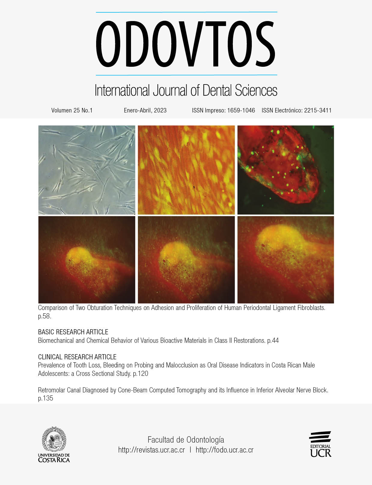
How to Cite
Abstract
The purpose of this research was the tomographic evaluation of the Mandibular Buccal Shelf (MBS) in orthodontic patients with different vertical growth pattern. An observational, descriptive, cross-sectional and retrospective study was conducted. Tomographic images of patients aged 14 to 40 years were observed and a database was formed with those that met the inclusion criteria. The sample size was 10 for each group according to vertical growth pattern (hypodivergent, normodivergent and hyperdivergent). Then four zones of frequent insertion of extralveolar mini-screws were selected in the MBS, taking as a reference the mesial and distal roots of the first and second mandibular molar. When comparing the characteristics of MBS between vertical growth patterns, between sexes and hemiarchs, no statistically significant differences were found. However, when the characteristics of MBS were compared according to the reference root, it was found that there were statistically significant differences. The vestibular area to the distal root of the second mandibular molar presented the highest values in terms of angulation, height and thickness. There are no significant differences in the bone characteristics of MBS according to vertical growth patterns, sexes or hemiarchs. Angulation, height and thickness progressively increase from the vestibular bone of the mesial root of the first mandibular molar to the distal root of the second molar.
References
Chang C., Huang C., Roberts W.E. 3D Cortical bone anatomy of the mandibular buccal shelf: a CBCT study to define sites for extra-alveolar bone screws to treat class III malocclusion. Int J Orthod Implan. 2016; 41:.74-82.
Jing Y., Han X., Guo Y., Li J., Bai D. Nonsurgical correction of a Class III malocclusion in an adult by miniscrew-assisted mandibular dentition distalization. Am J Orthod Dentofacial Orthop 2013; 143: 877-87.
Ramírez-Ossa D.M., Escobar-Correa N., Ramírez-Bustamante M.A., Agudelo-Suárez A.A. An umbrella review of the effectiveness of temporary anchorage devices and the factors that contribute to their success or failure. J Evid Based Dent Pract 2020; 20: 101402.
Marquezan M., Mattos C.T., Sant’Anna E.F., de Souza M.M., Maia L.C. Does cortical thickness influence the primary stability of miniscrews?: A systematic review and meta-analysis. Angle Orthod 2014; 84 (6): 1093-103.
Chen K., Cao Y. Class III malocclusion treated with distalization of the mandibular dentition with miniscrew anchorage: A 2-year follow-up. Am J Orthod Dentofacial Orthop. 2015; 148 (6): 1043-53.
Chang C., Liu S.S.Y., Roberts W.E. Primary failure rate for 1680 extra-alveolar mandibular buccal shelf mini-screws placed in movable mucosa or attached gingiva. Angle Orthod. 2015; 85 (6): 905-910.
Nucera R., Lo Giudice A., Bellocchio A.M., Spinuzza P., Caprioglio A., Perillo L., et al. Bone and cortical bone thickness of mandibular buccal shelf for mini-screw insertion in adults. Angle Orthod 2017; 87: 745-51.
Migliorati M., Benedicenti S., Signori A., Drago S., Barberis F., Tournier H., et al. Miniscrew design and bone characteristics: an experimental study of primary stability. Am J Orthod Dentofacial Orthop 2012; 142: 228-34.
Elshebiny T., Palomo J.M., Baumgaertel S. Anatomic assessment of the mandibular buccal shelf for miniscrew insertion in white patients. Am J Orthod Dentocial Orthop. 2018; 153 (4): 505-11.
Kapila S.D., Nervina J.M. CBCT in orthodontics: assessment of treatment outcomes and indications for its use. Dentomaxillofac Radiol. 2015; 44 (1): 20140282.
De Grauwe A., Ayaz I., Shujaat S., Dimitrov S., Gbadegbegnon L., Vande Vannet B., Jacobs R. CBCT in orthodontics: a systematic review on justification of CBCT in a paediatric population prior to orthodontic treatment. Eur J Orthod. 2019; 41 (4): 381-389.
Abdelkarim A. Cone-Beam Computed Tomography in Orthodontics. Dent. J. 2019; 7 (3): 89.
Sadek M.M., Sabet N.E., Hassan I.T. Three-dimensional mapping of cortical bone thickness in subjects with different vertical facial dimensions. Prog Orthod. 2016; 17 (1): 32.
Escobar-Correa N., Ramírez-Bustamante M.A., Sánchez-Uribe L.A., Upegui-Zea J.C., Vergara-Villarreal P., Ramírez-Ossa D.M. Evaluation of mandibular buccal shelf characteristics in the Colombian population: A cone-beam computed tomography study. Korean J Orthod. 2021 Jan 25; 51 (1): 23-31.
Aleluia R.B., Duplat C.B., Crusoé-Rebello I., Neves F.S. Assessment of the mandibular buccal shelf for orthodontic anchorage: Influence of side, gender and skeletal patterns. Orthod Craniofac Res. 2021; 24 Suppl 1: 83-91.
Rickets R.M., Roth R.H., Chaconnas S.J., Schulhof R.J., Engle G.A. Orthodontic diagnosis and planning. Denver: Rocky Mountain Data System; 1982.
Gregoret J., Tuber E., Escobar H., Matos-Da Fonseca A. Ortodoncia y cirugía ortognática, diagnostico y planificación. 2da edición. España: Editorial Publicaciones Médicas Barcelona; 1997.
Siriwat P.P., Jarabak J.R. Malocclusion and facial morphology is there a relationship? An epidemiologic study. Angle Orthod. 1985 Apr; 55 (2): 127-38.
García-Lallana A., Viteri-Ramírez G., Saiz-Mendiguren R., Broncano J., Dámaso J. Ergonomía del puesto de trabajo en radiología. Radiología. 2011; 53 (6): 507-15.
Echeverri S., Giraldo D., Lozano L., Mejía P.A.; Montoya L., Vásquez E.M. Síndrome de visión por computador: una revisión de sus causas y del potencial de prevención. Rev CES Salud Pública. 2012; 3 (2): 193-201.
Trivedi K., Jani B.K., Hirani S., Radia M.V. Comparative Evaluation of Cortical Bone Anatomy of Mandibular Buccal Shelf for Mini Implant Placement in Different Facial Divergence: A Cone Beam Computed Tomography Study. J Indian Orthod Soc. 2020; 54 (4): 325-331.
Gandhi V., Upadhyay M., Tadinada A., Yadav S. Variability associated with mandibular buccal shelf area width and height in subjects with different growth pattern, sex, and growth status. Am J Orthod Dentofacial Orthop. 2021 Jan; 159 (1): 59-70.
Parinyachaiphun S., Petdachai S., Chuenchompoonut V. Considerations for placement of mandibular buccal shelf orthodontic anchoring screw in Class III hyperdivergent and normodivergent subjects-A cone beam computed tomography study, Orthod Waves. 2018; 77 (1): 44-56.
Farnsworth D., Rossouw P.E., Ceen R.F., Buschang P.H. Cortical bone thickness at common miniscrew implant placement sites. Am J Orthod Dentofacial Orthop. 2011; 139: 495-503.
Kolge N.E., Patni V.J., Potnis S.S. Tomographic mapping of Buccal Shelf area for optimum placement of bone screws: A three-dimensional cone-beam computed tomography evaluation. APOS Trends Orthod 2019; 9 (4): 241-5.
Comments

This work is licensed under a Creative Commons Attribution-NonCommercial-ShareAlike 4.0 International License.
Copyright (c) 2022 CC-BY-NC-SA 4.0